Knee Muscle Anatomy Mri : Knee Muscle Anatomy Mri - knee anatomy | MRI knee coronal ...

View of the anatomical labels. Has stock or stock options held in conformis inc.; Want to learn more about it? Normal mr imaging anatomy of the knee. In relation to the pcl, the ligament of humphrey courses anterior, and the ligament of wrisberg courses posterior. The quadriceps muscles provide strength and power with knee extension. Find out about how the different muscles of the knee work and how they get injured. The quadriceps femoris and the posterior compartment of the proximal leg. Anatomy basic knee mri checklist. This mri knee sagittal cross sectional anatomy tool is.
Patellofemoral problems | the knee doc / 4, infrapatellar fat pad of hoffa. This long muscle flexes the knee. Serves as a paid consultant to or is an employee of conformis inc.; Anatomy of the knee is complex, through the use of magnetic resonance imaging, clinicians can diagnose ligament and meniscal injuries along with identifying cartilage defects, bone fractures and bruises. The knee joint is most significantly affected by two major muscle groups: Involved early gray = muscle: Want to learn more about it? Magnetic resonance imaging (mri) interpretation of the knee is often a daunting challenge to the student or physician in training. Anatomy basic knee mri checklist. Mri for evaluating knee pain in older patients:
The semimembranosus muscle is the largest of the posteromedial muscles continuing inferiorly to this level.
Use the checklist to quiz yourself. Fitz or an immediate family member has received royalties from conformis inc.; 4, infrapatellar fat pad of hoffa. Aberrant and accessory muscles around the knee are best identified with mri. Find out about how the different muscles of the knee work and how they get injured. Free cross sectional anatomy of the knee based on mri : General anatomy and musculoskeletal system. Knee anatomy wolfgang fitz, md jeffrey lange, md dr. The muscles of the knee joint are incredibly important. Although not dangerous, can cause pain if exposure increases 50. Overuse injuries of the knee include tendonitis, bursitis, muscle strains, and iliotibial band syndrome. Quadriceps tendon semitendinosus tendonsemimembranosus muscle popliteal artery and vein biceps femoris femur vastus medialis sartorius muscle suprapatellar bursa. Anatomy basic knee mri checklist.
Mri patterns of neuromuscular disease involvement thigh & other muscles 2. Tips to keep joints healthy. This long muscle flexes the knee. In the knee mri mastery courses, we give you everything you need in order to evaluate this joint. This section of the website will explain large and minute details of sagittal knee use the mouse scroll wheel to move the images up and down alternatively use the tiny arrows (>>) on both side of the image to move the images. Free cross sectional anatomy of the knee based on mri :

A coronal scan goes through the knee, front.
This long muscle flexes the knee. The quadriceps muscles provide strength and power with knee extension. This mri knee cross sectional anatomy tool is absolutely free to use. Use the checklist to quiz yourself. Click now to learn more about the bones, muscles, and soft tissues of these regions at leg and knee anatomy: The muscles of the knee joint are incredibly important. Has stock or stock options held in conformis inc.; Master leg and knee anatomy using our topic page. To begin, we use a coronal scan of a left knee. Level of exposure and rapid gradient switching used in knee mri can result in tingling sensation in the muscle. Click on the links to show each structure. Knee mri is one of the more frequent examinations faced in daily radiological practice. Patellofemoral problems | the knee doc / 4, infrapatellar fat pad of hoffa. Knee muscles need to have both good strength and flexibility. Mr arthrogram knee loose osteochondral lesion.
This mri knee cross sectional anatomy tool is absolutely free to use. Anatomy basic knee mri checklist. Articular surface of patella and femur, condyle, epicondyle and muscles (popliteus anatomy of the ankle and foot in mri: Has stock or stock options held in conformis inc.; Master leg and knee anatomy using our topic page. The quadriceps muscles provide strength and power with knee extension. 4, infrapatellar fat pad of hoffa. Learn about the muscles, tendons, bones, and ligaments that comprise the knee joint anatomy.

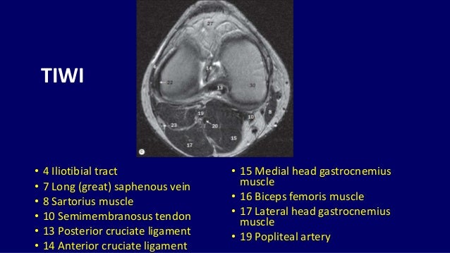
Quadriceps tendon semitendinosus tendonsemimembranosus muscle popliteal artery and vein biceps femoris femur vastus medialis sartorius muscle suprapatellar bursa.
The muscles of the knee include the quadriceps, hamstrings, and the muscles of the calf. View of the anatomical labels. Click now to learn more about the bones, muscles, and soft tissues of these regions at leg and knee anatomy: Musculoskeletal radiology south texas radiology group. The muscles of the knee joint are incredibly important. This long muscle flexes the knee. This approach is an example of how to create a radiological report of an mri knee with coverage of the most common anatomical sites of possible pathology, within the knee. Knee anatomy the orthopedic sports medicine institute in they. Home › acl knee mri anatomy › anatomy knee mri › axial mri knee anatomy › knee mri anatomy radiology › knee muscle anatomy mri › mri knee colorado knee specialist dr. On anatomical parts the user. Free cross sectional anatomy of the knee based on mri : 4, infrapatellar fat pad of hoffa. Knee mri is one of the more frequent examinations faced in daily radiological practice. Normal mri anatomy of the knee.

This mri knee cross sectional anatomy tool is absolutely free to use.

Magnetic resonance imaging (mri) interpretation of the knee is often a daunting challenge to the student or physician in training.

Click now to learn more about the bones, muscles, and soft tissues of these regions at leg and knee anatomy:

To begin, we use a coronal scan of a left knee.

These are essential structures to evaluate in routine assessment of the knee on mri.
 interpretation of the knee is often a daunting challenge to the student or physician in training.")
Involved early gray = muscle:

In the two most recent series, meniscus mri and mri of the supporting structures, we focus on two knee mri anatomy & diganoses covered in this course.

Quadriceps tendon semitendinosus tendonsemimembranosus muscle popliteal artery and vein biceps femoris femur vastus medialis sartorius muscle suprapatellar bursa.

The journal of musculoskeletal medicine.

Use the checklist to quiz yourself.

Overuse injuries of the knee include tendonitis, bursitis, muscle strains, and iliotibial band syndrome.

Has stock or stock options held in conformis inc.;

The knee joint is the junction of the thigh and leg.

Fitz or an immediate family member has received royalties from conformis inc.;

Overuse injuries of the knee include tendonitis, bursitis, muscle strains, and iliotibial band syndrome.

Knee anatomy wolfgang fitz, md jeffrey lange, md dr.

Knee muscles need to have both good strength and flexibility.

Mri for evaluating knee pain in older patients:

Articular surface of patella and femur, condyle, epicondyle and muscles (popliteus anatomy of the ankle and foot in mri:

This long muscle flexes the knee.

In relation to the pcl, the ligament of humphrey courses anterior, and the ligament of wrisberg courses posterior.

Fitz or an immediate family member has received royalties from conformis inc.;

Involved early gray = muscle:

Learn about the muscles, tendons, bones, and ligaments that comprise the knee joint anatomy.

Normal mr imaging anatomy of the knee.

Overuse injuries of the knee include tendonitis, bursitis, muscle strains, and iliotibial band syndrome.
:")
To begin, we use a coronal scan of a left knee.

Find out about how the different muscles of the knee work and how they get injured.

Tips to keep joints healthy.

Posting Komentar untuk "Knee Muscle Anatomy Mri : Knee Muscle Anatomy Mri - knee anatomy | MRI knee coronal ..."